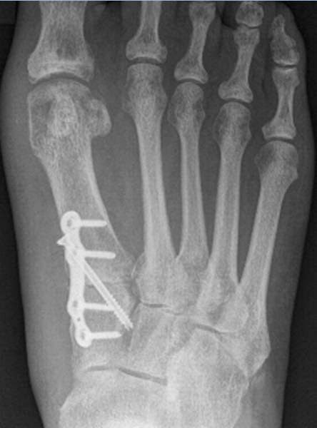
Most people are familiar with X-ray photography, and have seen pictures of broken bones, devices used to knit bones together, and pacemakers. Everyone has seen X-ray pictures. See below for a few examples (Click any image to enlarge).
Broken Leg
Broken Big toe, with plate and a drywall screw
Pacemaker with stimulating wires
X-rays are ideal for taking pictures of things that absorb X-rays, such as bone and metal. But if you need to look inside something made of metal... what then? X-rays won't make it through to the other side because they will be absorbed. Suppose we were to use neutrons instead?
Neutrons will go right through some metals and let you look at what is inside, because neutrons are absorbed by a different mechanism than X-rays. For example, if you wanted to image a rubber band inside of a lead brick, X-rays would be useless, but neutrons could do that. Let's look at a couple of examples of neutron radiography.
Below: An X-Ray image of a 35mm SLR camera: Note the lack of detail and large dark areas where metal has blocked the X-rays from reaching the film.
Below is a somewhat blurry image of an air compressor. Even with the blur, you can make out the crankshaft, connecting rod, wrist pin, valves, and even the roller bearings. Try doing that with X-rays.
The only problem with neutron radiography is that you can't use it on living tissue. Neutrons are pretty lethal, and they also get absorbed, making things radioactive. Additionally, people, being mostly water, are opaque to neutrons. Nevertheless, neutron radiography is a very useful tool for imaging the inside of metallic objects.
The way neutron radiography was accomplished at the facility is thus: A 12"x12" square aluminum dry tube was placed in the reactor pool, next to the core. The dry tube was 25 feet long so that it could reach the bottom of the pool and sit close to the reactor core.
The dry tube gave neutrons (and unwanted gamma radiation) a path to escape the core without being absorbed in water. The length of the tube served to "collimate" the neutrons - to have them traveling in the same direction as they exited the top. This collimation helps to sharpen the image.
At the top of the tube would be a thin aluminum plate, upon which the item to be imaged rested. Just above the item would be an aluminum cassette containing a large piece of photographic film. I no longer recall if it was special film or just ordinary X-ray film. The reactor power level required for this process wasn't terribly high - perhaps 5 watts for 10 minutes exposure time. However just 5 watts of unshielded radiation straight up from the reactor core is a nasty dose.
While performing neutron radiography, the receptionist was required to leave the building, since she was not a trained Radiation Worker. I wasn't keen on getting dosed with neutron and gamma either, and I was a lot closer to the dry tube than the receptionist desk. Fortunately, this was an infrequent operation, so it probably wasn't necessary to move the reactor control console to a separate room.
Neutron radiography is a very cool imaging technique, with some useful real-world applications.
The Air Force's F-111 Aardvark (as well as other aircraft) had aluminum honeycomb structure inside the wings for stiffness. It was found that corrosion was occurring within the honeycomb. It would have been incredibly tedious and expensive to peel back the skin of every F-111 wing at maintenance intervals to perform a visual inspection. Instead, the Air Force used neutron radiography with a new TRIGA reactor at McClellan Air Force Base in Sacramento.
The more you know!


No comments:
Post a Comment